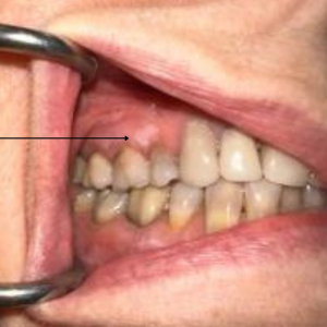
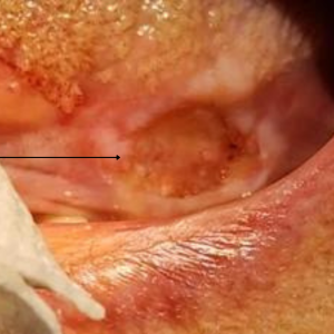
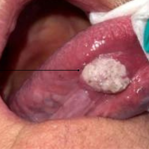
The inside of the mouth is normally lined with a special type of skin (mucosa) that is smooth and coral pink in color. Any alteration in this appearance could be a warning sign for a pathological process. The most serious of these is oral cancer. Oral mucosa changes can be detected on the lips, cheeks, palate, and gum tissue around the teeth, tongue, face and/or neck. Pain does not always occur with pathology, and curiously, is not often associated with oral cancer. However, any patient with facial and/or oral pain without an obvious cause or reason may also be at risk for oral cancer.
Historically the death rate associated with this cancer is particularly high, not because it is hard to detect or diagnose, but because the cancer is often discovered late in its development.
The National Cancer Institute’s SEER data indicate that when oral cancer is detected early, survival outcomes are improved and treatment-related health problems are reduced. Among healthcare professionals, your family dentist or oral and maxillofacial surgeon is in the best position to detect oral cancer during your routine dental examinations. If you are at high risk for oral cancer, you should see your general dentist or oral and maxillofacial surgeon for an annual exam.
See your oral and maxillofacial surgeon if you have any of these signs. If the oral and maxillofacial surgeon agrees that something looks suspicious, a biopsy may be recommended. A biopsy involves the removal of a piece of the suspicious tissue, which is then sent to a pathology laboratory for a microscopic examination that will accurately diagnose the problem. The biopsy report not only helps establish a diagnosis, but also enables the doctor to develop a specific plan of treatment.
Keep in mind that your mouth is one of your body’s most important early warning systems. Don’t ignore any suspicious lumps or sores. Should you discover something, make an appointment for a prompt examination. Early treatment may well be the key to complete recovery.